What is age-related macular degeneration?

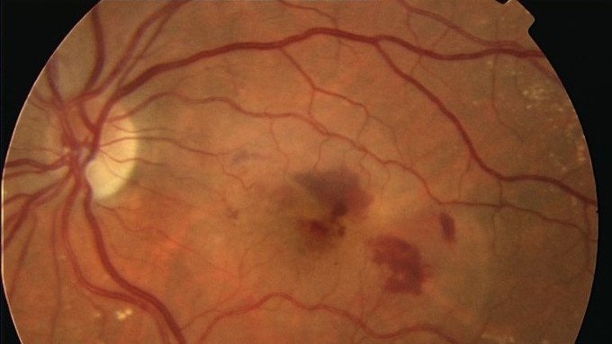
AMD is a degenerative disease of the macula, the central area of the retina, which causes progressive deterioration of the cells and the retinal pigment epithelium. It causes a loss of central vision.
There are two types:
- Dry AMD. Affecting 80% of patients, its progression is slow and gradual. The deposits that accumulate in the area cause atrophy of the macula, producing slow vision loss in the central area of the field of vision
- Wet AMD. Characterised by the growth of new blood vessels with very thin walls, resulting in the leakage of fluid and blood into the macula. Vision loss is rapid
What causes it?
AMD is a degenerative disease caused by the aging of the central area of the retina.
The main risk factors are:
- Age
- Smoking
- Genetic predisposition
- High blood pressure
How can it be prevented?
AMD cannot be prevented as it is associated with the aging process. However, since a higher incidence has been observed in smokers and people with a family history of the disease, certain measures can be taken.
The over-50s are recommended to eat a healthy diet, not smoke and have regular eye check-ups.
Symptoms
AMD sufferers progressively lose their central vision, causing difficulty in reading, writing, driving, sewing or other tasks that require precision.
Sufferers can have difficulty recognising people’s faces, but can walk without tripping and can maintain a certain level of independence. The disease usually starts in one eye, but eventually affects both.
As a result, sufferers tend not to be aware of visual problems unless they cover the good eye and notice that vision is distorted in the affected eye.
A simple test the over-50s can do once a week is to cover one eye and then the other, to see if straight lines formed by handrails or door frames appear distorted. If they do, the person should immediately visit their ophthalmologist.
Associated treatments
Wet AMD can be controlled with intravitreal antiangiogenic drugs, which have the function of slowing blood vessel growth.
For dry AMD, effective treatment has yet to be found, although administering antioxidant complexes can slow the disease. Studies are currently being carried out into genetic predisposition to AMD.
The aim in the near future is to identify people with a higher risk of suffering from the disease and to monitor them closely.
Specialists who treat this pathology
FAQs
Angiography is a technique used to delineate retinal or choroidal cases. Different contrasts are used, usually sodium fluorescein or indocyanine green. The scan is also useful for the diagnosis of other retinal diseases, such as pigment epithelium. In general, angiography is used to study many retinal diseases and their diagnosis.
It is a diagnostic technique to determine pathological and abnormal structures in the blood vessels and the different layers of the retina. It can be used in cases of macular degeneration, diabetic retinopathy, vasculopathy and many other macular disorders.
It is not counterproductive for any eye treatment.
It is a grid of straight horizontal and vertical lines with a central point, used as a tool to detect visual disturbances in patients with scotoma or other anomalies.
Age-related macular degeneration is one of the greatest challenges in ophthalmology today. We know that there are two types: the dry form and the wet form. The dry form is experienced by patients who are slowly losing their vision. It has been demonstrated that treatment with antioxidants and vitamins can reduce vision loss, although the slowing down of the process is not particularly spectacular. The wet form, which is so called because fluid is produced in the macula, is the most destructive, and current treatment involves the combining of photodynamic therapy, which started to be used some years ago, with other treatments, which has produced more positive results. Each year, new possibilities appear, which help in the fight against this disease.
Yes, it is an emergency, but relatively speaking, as it is possible to wait 3 or 4 days. It is necessary to examine the eye, because the symptom could indicate the onset of decompensation, which can cause severe loss of vision. This distortion is sometimes not due to decompensation, but it always needs to be confirmed.
IMO Institute of Ocular Microsurgery
Josep María Lladó, 3
08035 Barcelona
Phone: (+34) 934 000 700
E-mail: international@imo.es
See map on Google Maps
By car
GPS navigator coordinates:
41º 24’ 38” N – 02º 07’ 29” E
Exit 7 of the Ronda de Dalt (mountain side). The clinic has a car park with more than 200 parking spaces.
By bus
Autobus H2: Rotonda de Bellesguard, parada 1540
Autobus 196: Josep Maria Lladó-Bellesguard, parada 3191
Autobuses H2, 123, 196: Ronda de Dalt – Bellesguard, parada 0071
How to arrive at IMO from:
IMO Madrid
C/ Valle de Pinares Llanos, 3
28035 Madrid
Phone: (+34) 910 783 783
See map in Google Maps
Public transport
Metro Lacoma (líne 7)
Autobuses:
- Lines 49 & 64, stop “Senda del Infante”
- Line N21, stop “Metro Lacoma”
Timetables
Patient care:
Monday to Friday, 8 a.m. to 9 p.m.
IMO Andorra
Av. de les Nacions Unides, 17
AD700 Escaldes-Engordany, Andorra
Phone: (+376) 688 55 44
See map in Google Maps
IMO Manresa
C/ Carrasco i Formiguera, 33 (Baixos)
08242 – Manresa
Tel: (+34) 938 749 160
See map in Google Maps
Public transport
FGC. Line R5 & R50 direction Manresa. Station/Stop: Baixador de Manresa
Timetables
Monday to Friday, 09:00 A.M – 07:00 PM

