What is autofluorescence?
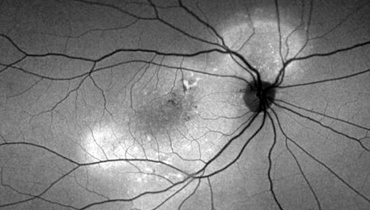
Autofluorescence is a technique that allows the metabolic changes that occur in the retinal pigment epithelium (RPE), an essential layer to keep photoreceptor cells in good health, to be viewed in great detail.
The information provided by this test helps the ophthalmologist to study the origin and evolution of certain retinal pathologies to be able to make a more precise diagnosis and select the most appropriate treatment in each case.
What does it consist of?

Autofluorescence leverages the fluorescent properties of a metabolic indicator called lipofuscin, which is present in the fundus of the eye as a result of light stimulation of the cellular pigments of the retinal pigment epithelium.
Thanks to the development of diagnostic imaging technologies, the study of lipofuscin accumulation patterns can be performed in a non-invasive way for the patient, while viewing its deposits in vivo, since an increase or decrease in certain areas reflects pathological alterations.
Wide-field systems are very useful to reach even the most peripheral areas, which require a single image capture to view 200° of the retina (vs 30 – 45° of the central field).
How is it performed?
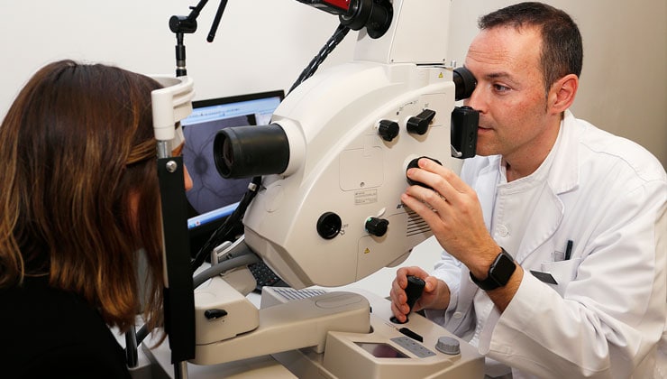
This test is performed in about 5 minutes. To do so, it is necessary to dilate the pupil with drops (mydriatic eye drops) that take effect in about 15 minutes. As a result of the increased pupil diameter, the patient experiences blurred vision and glare, although these symptoms disappear after a few hours.
In what cases is it used?
Autofluorescence is indicated for patients with retinal disorders that affect the pigment epithelium, the main ones being retinal dystrophies and AMD, in addition to drug toxicity, posterior uveitis or some intraocular tumours, among others.
This technique is especially useful in unexplained visual acuity losses and in hereditary retinal dystrophies, such as retinitis pigmentosa or Stargardt disease. The processes involved have been shown to be related to alterations in this layer, which can help identify the specific type of dystrophy and guide genetic counselling.
Associated pathologies
IMO Institute of Ocular Microsurgery
Josep María Lladó, 3
08035 Barcelona
Phone: (+34) 934 000 700
E-mail: international@imo.es
See map on Google Maps
By car
GPS navigator coordinates:
41º 24’ 38” N – 02º 07’ 29” E
Exit 7 of the Ronda de Dalt (mountain side). The clinic has a car park with more than 200 parking spaces.
By bus
Autobus H2: Rotonda de Bellesguard, parada 1540
Autobus 196: Josep Maria Lladó-Bellesguard, parada 3191
Autobuses H2, 123, 196: Ronda de Dalt – Bellesguard, parada 0071
How to arrive at IMO from:
IMO Madrid
C/ Valle de Pinares Llanos, 3
28035 Madrid
Phone: (+34) 910 783 783
See map in Google Maps
Public transport
Metro Lacoma (líne 7)
Autobuses:
- Lines 49 & 64, stop “Senda del Infante”
- Line N21, stop “Metro Lacoma”
Timetables
Patient care:
Monday to Friday, 8 a.m. to 9 p.m.
IMO Andorra
Av. de les Nacions Unides, 17
AD700 Escaldes-Engordany, Andorra
Phone: (+376) 688 55 44
See map in Google Maps
IMO Manresa
C/ Carrasco i Formiguera, 33 (Baixos)
08242 – Manresa
Tel: (+34) 938 749 160
See map in Google Maps
Public transport
FGC. Line R5 & R50 direction Manresa. Station/Stop: Baixador de Manresa
Timetables
Monday to Friday, 09:00 A.M – 07:00 PM


