The cornea

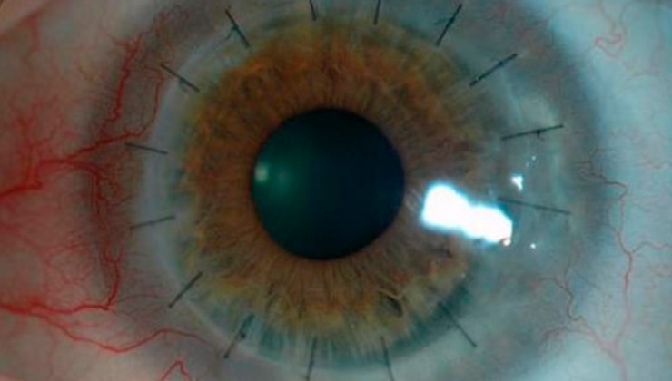
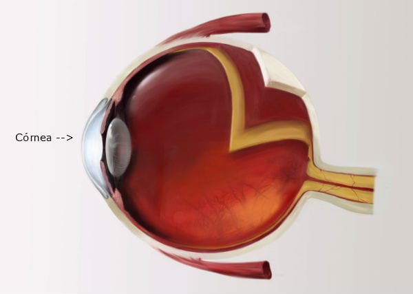
The cornea is located in the front part of the eye and is essentially the eye’s window. Under normal conditions, the cornea is completely transparent due to the large number of fibres that form it and the absence of blood vessels. This feature, together with the refractive power of the cornea and the lens, allows the images we see to be projected onto the retina, which is the part of the eye that actually “sees” the images.
What is Peters anomaly?

Peters anomaly is a congenital error in the development of the eye that occurs between 10 and 16 weeks of gestation. It can affect one or both eyes and usually cannot be detected through prenatal screening.
It tends to be a one-off abnormality in a family, although sometimes it affects more than one family member. Genetic investigation of the family is important, since the genetic error (mutation) that causes it can be multiple; in other words, it does not only affect the eyes, but is also associated with mutations in other genes that affect other organs or systems, such as heart disease, deafness, learning difficulties, cleft lips and cleft palates. When this occurs, it is known as Peters-plus syndrome.
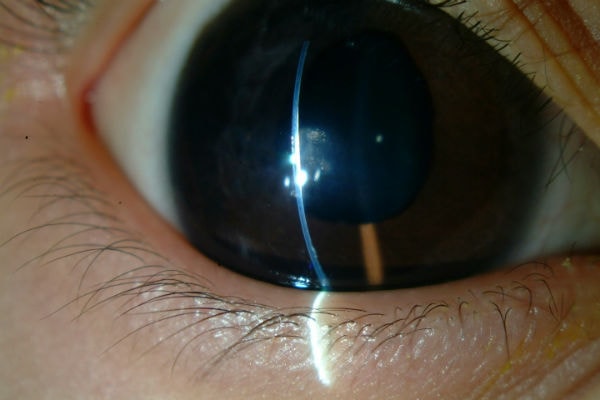
In this condition, the central part of the cornea is opaque (i.e. white). The fact that the central part of the cornea is not transparent always results in a loss of vision, whose severity increases with the degree of opacity If the abnormality affects both eyes, the child cannot recognise objects, although he or she may be able to perceive masses and recognise colours.
Although opacity tends to be the most obvious sign in these patients, corneal alteration occurs alongside other developmental abnormalities in the front part of the eye. It may also be associated with an increased risk of glaucoma (increased ocular pressure that can damage the optic nerve), nystagmus nystagmus (involuntary eye movements), cataracts (a loss of transparency in the lens) and retinal detachment.
The condition varies in severity. In the most serious cases of underdevelopment, the eye is smaller (microphthalmia) and rudimentary.
How is it diagnosed?
The condition is usually detected by a paediatrician straight after birth, although Peters anomaly must be diagnosed by an ophthalmologist. It is important that a diagnosis be made as early as possible; sometimes a thorough eye examination under general anaesthetic is required.
Although central corneal opacity usually improves spontaneously during the first months of life, it is very difficult to predict the degree of vision the child will have.
Symptoms
The central part of the cornea is opaque (i.e. white).
If the abnormality affects both eyes, the child cannot recognise objects, although he or she may be able to perceive masses and recognise colours.
Although opacity tends to be the most obvious sign in these patients, corneal alteration occurs alongside other developmental abnormalities in the front part of the eye. It may also be associated with an increased risk of glaucoma, nystagmus, cataracts and retinal detachment.
In the most serious cases of underdevelopment, the eye is smaller (microphthalmia) and rudimentary.
Associated treatments
In cases where both eyes are affected, the only way to improve visual acuity is through corneal transplantation. Surgery is carried out on an outpatient basis under general anaesthetic.
If the patient also presents glaucoma, this must be treated. Sometimes treatment with eye drops is enough to normalise the pressure, but at other times, surgery is required.
Lastly, if the child presents acute photophobia, wearing sunglasses may help.
Specialists who treat this pathology
IMO Institute of Ocular Microsurgery
Josep María Lladó, 3
08035 Barcelona
Phone: (+34) 934 000 700
E-mail: international@imo.es
See map on Google Maps
By car
GPS navigator coordinates:
41º 24’ 38” N – 02º 07’ 29” E
Exit 7 of the Ronda de Dalt (mountain side). The clinic has a car park with more than 200 parking spaces.
By bus
Autobus H2: Rotonda de Bellesguard, parada 1540
Autobus 196: Josep Maria Lladó-Bellesguard, parada 3191
Autobuses H2, 123, 196: Ronda de Dalt – Bellesguard, parada 0071
How to arrive at IMO from:
IMO Madrid
C/ Valle de Pinares Llanos, 3
28035 Madrid
Phone: (+34) 910 783 783
See map in Google Maps
Public transport
Metro Lacoma (líne 7)
Autobuses:
- Lines 49 & 64, stop “Senda del Infante”
- Line N21, stop “Metro Lacoma”
Timetables
Patient care:
Monday to Friday, 8 a.m. to 9 p.m.
IMO Andorra
Av. de les Nacions Unides, 17
AD700 Escaldes-Engordany, Andorra
Phone: (+376) 688 55 44
See map in Google Maps
IMO Manresa
C/ Carrasco i Formiguera, 33 (Baixos)
08242 – Manresa
Tel: (+34) 938 749 160
See map in Google Maps
Public transport
FGC. Line R5 & R50 direction Manresa. Station/Stop: Baixador de Manresa
Timetables
Monday to Friday, 09:00 A.M – 07:00 PM


