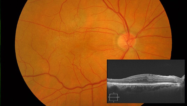
What is MEM?

Macular epiretinal membrane (MEM) means tissue grows over the surface of the retina in the macular area that can contract and cause a loss of sight and image distortion.
What causes it?
It is caused by some cells being deposited over the macula that usually come from the retina, but can come from the layers located below the retina, such as the pigment epithelium.
These cells secrete collagen, forming a mesh, and can then apply traction to the collagen. Since this tissue is attached to the retina, when it contracts the retina also contracts and warps.
How can it be prevented?
These membranes most commonly appear in patients over the age of 50, but can also occur at any age. They are sometimes a result of a retinal hole, which enables cells to pass through the retina from the sub-retinal area.
It is therefore very important that before symptoms appear that could imply a retinal hole, such as floaters or flashing lights, you see your ophthalmologist to have these holes immediately repaired. In this way you can also avoid retinal detachment.
It can also appear after ocular surgery, hence post-operative monitoring is very important.
Symptoms
The most common symptoms are loss of sight, blurred vision or image distortion.
Lines may be seen distorted and numbers and letters can appear to jump out of line.
Associated treatments
It is very important to monitor that a retinal detachment is not being formed by examination in the post-operative period and seeing an ophthalmologist if you suffer any of these symptoms.
The most suitable treatment for MEM is a vitrectomy.
Specialists who treat this pathology
FAQs
Angiography is a technique used to delineate retinal or choroidal cases. Different contrasts are used, usually sodium fluorescein or indocyanine green. The scan is also useful for the diagnosis of other retinal diseases, such as pigment epithelium. In general, angiography is used to study many retinal diseases and their diagnosis.
Indocyanine green angiography is a technique used in some cases of AMD and serves to define the neovessels and, occasionally, to diagnose other diseases. Fluorescein angiography is the standard technique for studying blood vessel diseases and the retina in general.
It is a diagnostic technique to determine pathological and abnormal structures in the blood vessels and the different layers of the retina. It can be used in cases of macular degeneration, diabetic retinopathy, vasculopathy and many other macular disorders.
It is not counterproductive for any eye treatment.
Yes, after any eye operation, although it is advisable to wait several days for the scars to heal. It normally takes ten to twelve days to return to a normal life. It is always best to consult the doctor to find out about the risk factors.
IMO Institute of Ocular Microsurgery
Josep María Lladó, 3
08035 Barcelona
Phone: (+34) 934 000 700
E-mail: international@imo.es
See map on Google Maps
By car
GPS navigator coordinates:
41º 24’ 38” N – 02º 07’ 29” E
Exit 7 of the Ronda de Dalt (mountain side). The clinic has a car park with more than 200 parking spaces.
By bus
Autobus H2: Rotonda de Bellesguard, parada 1540
Autobus 196: Josep Maria Lladó-Bellesguard, parada 3191
Autobuses H2, 123, 196: Ronda de Dalt – Bellesguard, parada 0071
How to arrive at IMO from:
IMO Madrid
C/ Valle de Pinares Llanos, 3
28035 Madrid
Phone: (+34) 910 783 783
See map in Google Maps
Public transport
Metro Lacoma (líne 7)
Autobuses:
- Lines 49 & 64, stop “Senda del Infante”
- Line N21, stop “Metro Lacoma”
Timetables
Patient care:
Monday to Friday, 8 a.m. to 9 p.m.
IMO Andorra
Av. de les Nacions Unides, 17
AD700 Escaldes-Engordany, Andorra
Phone: (+376) 688 55 44
See map in Google Maps
IMO Manresa
C/ Carrasco i Formiguera, 33 (Baixos)
08242 – Manresa
Tel: (+34) 938 749 160
See map in Google Maps
Public transport
FGC. Line R5 & R50 direction Manresa. Station/Stop: Baixador de Manresa
Timetables
Monday to Friday, 09:00 A.M – 07:00 PM


